

Giant lung cryptococcoma in immunocompetent patient: case report
Mestre-Orozco, Laura1; Vicuña-González, Rosa María1; Domínguez-Sosa, Freddy Rafael1; López-Valdés, Julio César1
2022, Number 2
2022; 81 (2)


Mestre-Orozco, Laura1; Vicuña-González, Rosa María1; Domínguez-Sosa, Freddy Rafael1; López-Valdés, Julio César1
ABSTRACT
Lung cryptococcosis is a rare entity whose epidemiology hasn't been entirely reported in Mexico. It is frequently found as a complication of HIV or other cases of immunosuppression. We present a case of a young immunocompetent man who debuted with neurological manifestations and was later found to have a giant lung cryptococcoma. This is a particularly interesting case because although the clinical manifestations were normal, the patient was not immunocompromised and the lung damage was important with later development of meningitis that led to complications and a lung resection.KEYWORDS
cryptococcoma, giant, lung, immunocompetent.Introduction
Pulmonary cryptococcosis is a rare entity, whose epidemiology in Mexico is not fully described; however, according to data provided by Carrada BT et al.1 in Mexico the fungus was isolated in 20.7% of pigeon droppings samples in urban areas; in addition, they mention that in 1970 only 25 cases of cryptococcosis were registered in the hospitals of the Distrito Federal (now Mexico City).
On the other hand, they mention 13 cases confirmed after post mortem histopathological study out of a total of 22,088 studies (0.59 diagnoses per 1,000). However, it should be remembered that it frequently occurs as a complication of HIV infection or other immunosuppression (leukemia, use of glucocorticoids, among others), thus isolated cases in immunocompetent patients are considered even scarcer.2 However, the clinical picture in immunocompetent patients usually has a pulmonary onset with subsequent extension to the central nervous system (meningitis).3 The following is the case of a young, immunocompetent man who presented with an incipient clinical picture with neurological manifestations and in whom a giant pulmonary cryptococcoma was evidenced.
Case presentation
A 23-year-old man, originally from Oaxaca and resident of Monterrey, Nuevo León; he confirmed coexistence with animals such as urban pigeons, with no other relevant history. Four days after the application of the second dose of vaccine against the SARS-CoV-2 virus, he started with moderate to severe oppressive headache, which did not present irradiation and did not improve with analgesics; after two days of evolution, gait instability was added, as well as visual hallucinations and tonic-clonic convulsive crises.
He went to the hospital unit where a simple cranial tomography was performed, which showed the presence of multiple circumscribed lesions of heterogeneous distribution in both hemispheres of the brain. Laboratory studies were performed, consisting of serological tests for antibodies against HIV, HBV and HCV, which were negative. Treatment was started with amphotericin B deoxycholate 100 mg every 24 hours intravenously and fluconazole 100 mg every 12 hours orally, with clinical improvement after administration; however, he presented progressive decrease in visual acuity, as well as repeated febrile symptoms.
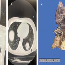
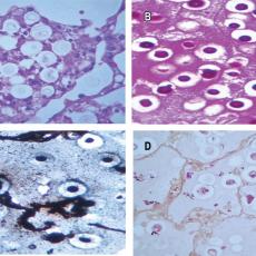
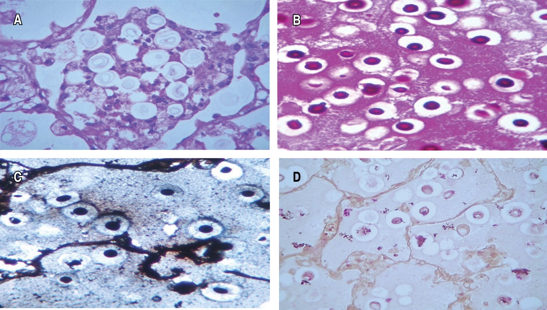
A simple chest CT scan of the lung was performed (Figure 1A and 1B), which showed a left lower lobar lesion. After three days without improvement, posterolateral thoracotomy and left lower lobectomy were performed, showing whitish-yellow consolidation of 10 × 10 cm in the lower lobe, upper lobe without alterations (Figure 1C). PAS, Grocott and mucicarmin stains were performed, showing abundant Cryptococcus throughout the lung parenchyma (Figure 2).
The patient was discharged after 27 days of stay, with outpatient management and antifungal regimen until further evaluation.
Discussion
The aforementioned case represents a common example of pulmonary cryptococcosis, whose clinical onset is usually incipient until presenting systemic manifestations associated with the infection; however, its particularity is related to the fact that even without presenting immunocompromise, pulmonary involvement was significant and there was progression to meningitis with associated neurological deficit that led to complications and pulmonary resection. Although it was not possible to determine the association with the second dose of the vaccine, in some way the evidence presented and the abrupt response after its application may imply an association related to the immunosuppression-immunocompetence status.
On the other hand, it has been described that when it is cryptococcosis isolated to the lung, the diagnosis is relatively good, however, this is drastically associated with a bad evolution when it has extrapulmonary extension, as in our case, and it is reported with up to 55% mortality.4 The dimensions of the exposed cryptococcoma are comparable to the largest pulmonary cryptococcoma published to date: the case of Menon A. et al.5 of 14 × 13 × 0.5 cm in a 48-year-old immunocompetent male patient without extrapulmonary extension. In that case, the patient had a two-year evolution time until he underwent a right upper lobectomy with subsequent resolution of the condition, and at eight-month follow-up showed no recurrence.
Due to the low incidence of cryptococcosis in Mexico, there is no consensus on the therapeutic regimen.6 Immunocompetent patients with isolated pulmonary cryptococcosis have an excellent prognosis without the need for antifungal therapy or surgical resection, which is why they are often left for observation only.7 B. Saag et al.8 recommend only initiating antifungal therapy if the patient is symptomatic. Nadrous HF et al.9 follow these recommendations, as well as indicating treatment when extrapulmonary extension is present as in the case of the patient presented here. Surgical treatment should be considered for cases with vital organ obstruction, persistent radiographic abnormalities, or cases refractory to treatment after one month. In this case, surgical treatment was decided because of continued neurological deterioration with no response to antifungal therapy.
Conclusions
This case represents a common example of pulmonary cryptococcosis, whose particularity lies in its presentation in a patient without immunocompromise. Even so, both pulmonary and neurological involvement was important. Although it was not possible to determine a relationship with the application of the second dose of the vaccine, the evidence presented and the abrupt start after its application may imply an association with the immunosuppression-immunocompromised state.
AFILIACIONES
1Hospital Central Sur de Alta Especialidad PEMEX. Mexico.Conflict of interests: the authors declare that they have no conflict of interests.
REFERENCES